


Introduction
Since the dawn of Vedic period (3500-16 BC), as described in Ayurvedic and Unani literature, medicinal plants formed the basis of all developments in medical sciences (Pattanayak et al., 2010). In the alternative and complementary medical practice, aromatic and medicinal plant parts such as shoots, leaves, roots, and seeds are the major sources of therapeutic agents (Jiang et al., 2015; Sun et al., 2014). The antioxidant potential of these plants has been attributed to the abundance of bio-active compounds also known as secondary metabolites such as alkaloids, flavonoids, phenols, and saponins, which are chemically complex and constitute a diverse mixture of molecules (Wu et al., 2016). These components suppress redox reactions of free radicals in the biological system and thus prevent the human body from suffering due to various ailments (Gao et al., 2012).
Among all medicinal plants, aromatic herbal plants are the major sources of bioactive agents useful in medicine. Of these plants, Ocimum spp., also known as Tulsi, holy basil, Queen of plants, and Mother Medicine of nature, has received extensive attention because of its diverse medicinal values (Beatović et al., 2013). There are approximately 160 species identified under the genus Ocimum that belong to the family Lamiaceae and are cultivated worldwide in the temperate regions (Hussain et al., 2017). In several countries such as Australia, East Asia, Europe, and USA, Ocimum spp. have been cultivated for commercial uses and are extensively used in food and perfume industries (Mandave et al., 2014). In India, this herb is worshipped in almost every home. It is most valuable and holistic plant used in traditional medicine because of its therapeutic properties (Singh et al., 2010). Traditionally, aqueous extracts of Ocimum leaves (dried powder or fresh) are used in herbal tea or mixed with honey to enhance its medical potential (Pattanayak et al., 2010). Tulsi oil extracted from this plant shows diverse biological activities such as analgesic, antiemetic, hypoglycemic, immune-booster, antibacterial, stress reliever, and expectorant (Yamani et al., 2016). The antioxidant potential and diverse biological activities of Ocimum spp. are based on the diverse range of secondary metabolites such as phenolics and flavonoids (Naquvi et al., 2012). Thus, the present study aimed to perform solvent-dependent extraction of secondary metabolites from the leaves of Ocimum basilicum L. (Green tulsi), Ocimum gratissimum L. (Jungli tulsi), and Ocimum tenuiflorum (Black tulsi) and to evaluate their total antioxidant activity and antidiabetic and anti-inflammatory activities.
Materials and methods
Plant material
Samplings of two varieties of tulsi, namely O. basilicum L. (Green tulsi) and O. tenuiflorum (Black tulsi), were purchased from local nursery. The O. gratissimum L. (Jungli tulsi) sample was a kind gift from Indian Institute of Integrative Medicine (CSIR-IIIM), Jammu, India. The plant species were verified by Dr. Upma from Botany Department at CSIR-IIIM. The plants were cultivated at CSIR-AROMA nursery in the campus and grown under the natural conditions of Jalandhar, India, located at 30°33ˊ North and 71°31ˊ East. The sampling was performed in April 2020. The city has a humid subtropical climate with cool winters and long, hot summers. The climate is dry on the whole. The mean max/min temperatures were 35°C/13°C, and the day length was 12 h 50 min. Fresh leaves of all Ocimum spp. were harvested, washed with distilled water, and shade dried. The dried leaves were milled to powder by using a coffee blender. For further processing, the powdered material was stored in food-grade aluminum foils at −20°C.
Preparation of plant extracts
Four different solvents with increasing polarity, namely acetone/water (8 : 2 v/v), ethanol/water (9 : 1, v/v), methanol/water (8 : 2, v/v), and water, were used to prepare plant extracts of all Ocimum species. Plant extracts were obtained by magnetic stirring of 3.0 g of dried powder with 30 ml of each solvent for 30 min at room temperature. The extracts were kept in dark for 24 h at 4°C.
Phytochemical analyses
The presence of various phytochemicals in tulsi leaf extracts was determined following the protocols of Sharma et al. (2021).
Total phenolic content
The total phenolic content of the Ocimum leaf extracts was measured by the Folin-Ciocalteu method. Briefly, 50 μl of samples was made up to 3 ml with double-distilled water, mixed properly with 0.5 ml of 1 : 10 Folin-Ciocalteu reagent, and incubated for 3 min. After incubation, 2 ml of 20% (w/v) sodium carbonate was added to each sample. The resulting solution was incubated for 60 min in dark before absorbance reading at 650 nm. Gallic acid (5–50 μg/ml) was used for the calibration curve. The results are expressed as mg of gallic acid equivalent (GAE) per gram (g) of dry weight (DW) based on the standard curve equation y = 0.0123x + 0.1476, R 2= 0.8835. All the sample were analyzed in triplicate.
Total flavonoid content analysis
The total flavonoid content of Ocimum leaf extracts was measured by the aluminum chloride colorimetric method. In brief, 10 μl of samples was made up to 1 ml with methanol and then mixed with 4 ml of doubledistilled water. Next, 0.3 ml of 5% NaNO2 was added to each sample, and the mixtures were incubated for 5 min. After incubation, 0.3 ml of 10% AlCl3 was added, and the mixtures were allowed to stand for 6 min. Next, 2 ml of 1 mol/l NaOH solution was added to each sample, the final volume of the reaction mixtures was brought to 10 ml with double-distilled water, and the mixtures were incubated for 15 min in dark. After incubation, the absorbance was measured at 510 nm. Rutin (50–500 μg/ml) was used for the calibration curve. The results are expressed as mg of rutin equivalent per g of DW using the standard curve equation y = 0.0005x, R2 = 0.7683. All the samples were analyzed in triplicate.
Total condensed tannin analysis
Condensed tannin (proanthocyanidins) content was determined according to the method of Sun et al. (2014). The reaction mixtures were prepared as follows: 50 μl of tulsi samples, 3.5 ml of 5% vanillin solution, and 2.5 ml of concentrated HCl were mixed together. The mixtures were incubated for 15 min at room temperature, and the absorption was measured at 500 nm by using methanol as a blank. The results are expressed as mg of ascorbic acid equivalent per g of DW using the standard curve equation y = 0.0006x, R 2 = 0.8683. All samples were analyzed in triplicate.
Spectral analysis of Ocimum extracts
Ocimum extracts were analyzed by UV-VIS spectro-photometry (Labtronics) in the range of 200–400 nm. UV spectra were recorded and analyzed. A Fourier transform infrared (FT-IR) study was conducted to identify the functional groups in the active constituents of Ocimum extracts. For this purpose, small amounts (approximately 10 μl) of Ocimum leaf extracts were taken. An IR spectrum was recorded at the wavelength spectrum from 4000 to 400 cm−1 by using an FT-IR spectrophotometer (Perkin Elmer, USA). Fluorescence analysis of the samples was performed using a fluorescent spectro-photometer (FL6500, Perkin Elmer, USA). For this purpose, 2 ml of samples was taken in quartz cuvettes and subjected to excitation at 380 nm.
Anti-inflammatory activity (protein denaturation) of Ocimum extracts
Protein denaturation assay was performed according to the protocol of Sharma et al. (2021). The reaction mixture consisted of 0.4 ml of 1% bovine serum albumin (BSA), 4.78 ml of phosphate-buffered saline (PBS with pH 6.4), and Ocimum extracts. The reaction mixtures were incubated in a water bath at 37°C for 15 min. The reaction mixtures were then heated at 70°C for 5 min and immediately cooled down. After cooling, 1 ml of each mixture was subjected to fluorescent spectroscopy. The excitation wavelength was 280 nm, and the fluorescence emission spectrum was noted in the wavelength range of 300–400 nm. The experiments were performed at room temperature (~30°C).
DPPH radical scavenging activity of Ocimum extracts
The free radical scavenging activity of Ocimum extracts was determined by the DPPH assay (Sharma et al., 2021). DPPH solution (2.8 ml) (4 mg DPPH in 100 ml of 82% MtOH) was added to Ocimum extracts (100 μl) and incubated at room temperature (25–27°C) in dark for 1 h. The absorbance values of the mixtures were then measured at 517 nm. The mixture of 0.2 ml of pure methanol and 2.8 ml of 82% methanol was used as blank, whereas 2.8 ml of DPHH and 0.2 ml of pure methanol was used as a control. Ascorbic acid was used as a positive control. The entire test was conducted in triplicates. The percentage of inhibition was determined using the formula:
Total antioxidant capacity of Ocimum extracts
Ocimum extract samples (100 μl) were added to 1 ml of reagent solution (0.6 mM H2SO4, 28 mM Na3PO4, and 4 mM ammonium molybdate). The tubes were incubated at 95°C for 90 min, followed by cooling to room temperature. The absorbance of each solution was measured at 695 nm against blank. Antioxidant capacity was expressed as an ascorbic acid equivalent per g of DW, y = 0.125x + 0.013, R 2 = 0.976. The entire sample was analyzed in triplicate.
Alpha-amylase inhibitory assay of Ocimum extracts
A total of 250 μl of Ocimum extract samples was placed in tubes, and to each tube, 125 μl of 0.02 M sodium phosphate buffer (pH 6.9) with α-amylase (5 mg/ml) was added. Sodium phosphate buffer (pH 6.9) (500 μl, 0.02 M) was then added to each tube, and the mixtures were incubated at 25°C for 20 min. Next, 600 μl of starch solution (2%) in 0.03 M phosphate buffer (pH 6.9) was added and incubated further at 25°C for 10 min. Dinitrosalicyclic acid (500 μl) was added to stop the reaction. The reaction tubes were incubated in a water bath at 100°C for 10 min. Subsequently, 6 ml of water was added, and optical density (OD) was measured at 540 nm. The α-amylase inhibitory activity was calculated as follows:
For control, the sample was replaced with water.
Results and discussion
Antioxidant analysis of Ocimum extracts
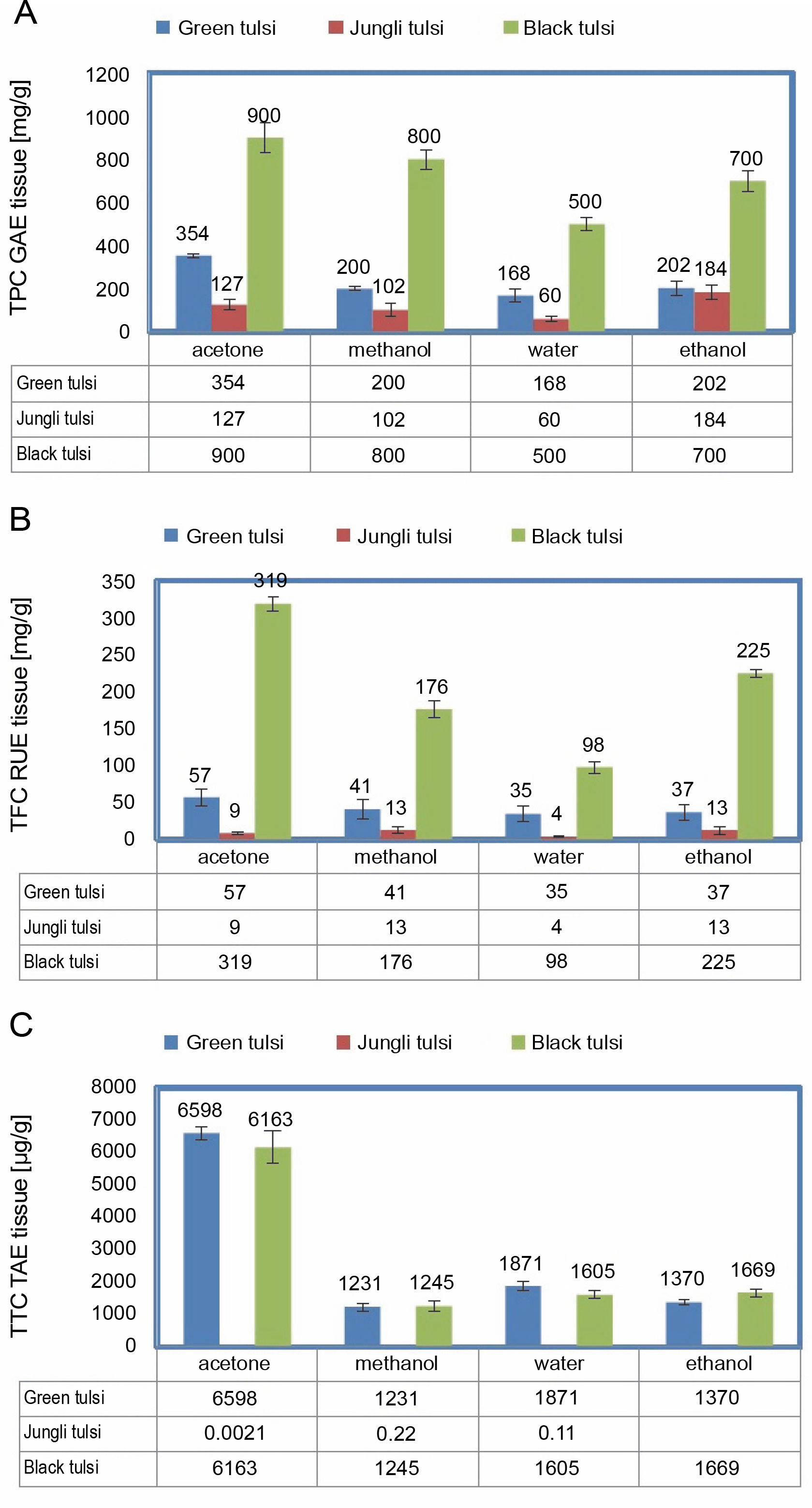
Ocimum spp. are extensively used as therapeutic agents in traditional medicine as they contain a diverse range of bioactive components (Naquvi et al., 2012). The extraction of bioactive molecules is highly dependent on the type (polarity) of solvent used (Medini et al., 2014). In the present study, acetone provided the maximum yield of phenolics in the range of 127 to 900 mg GAE/g DW in all Ocimum species, followed by methanol (102–800 mg GAE/g DW), ethanol (184–700 mg GAE/g DW), and water (60–500 mg GAE/g DW) (Fig. 1A). In acetone as the solvent, among all Ocimum spp., black tulsi showed the highest content of phenolics (900 mg GAE/g DW), followed by green tulsi (354 mg GAE/g DW) and jungli tulsi (127 mg GAE/g DW). A nearly similar trend was also observed for flavonoids (Fig. 1B). In the present study, acetone provided the maximum yield of flavonoids in the range of 9 to 319 mg rutin equivalent (RU)/g DW in all Ocimum species, followed by ethanol (13–225 mg RU/g DW), methanol (13–176 mg RU/g DW), and water (4–98 mg RU/g DW) (Fig. 1B). In acetone as the solvent, among all Ocimum spp., black tulsi showed the highest content of flavonoids of 319 mg RU/g DW, followed by green tulsi (57 mg RU/g DW) and jungli tulsi (9 mg RU/g DW). Regarding the content of total condensed tannins, acetone provided the maximum yield in the range of 2.1–659 mg tannic acid equivalent (TAE)/g DW in all Ocimum species, followed by ethanol (137–166 mg TAE/g DW), methanol (22–124 mg TAE/g DW), and water (11–187 mg TAE/g DW) (Fig. 1C). Among the three Ocimum spp., green tulsi showed the highest content of tannins of 659 mg TAE/g DW, followed by black tulsi (616 mg TAE/g DW) and jungli tulsi (2.1 mg TAE/g DW). This finding was consistent with an earlier study of Medini et al. (2014) on Limonium delicatulum, wherein acetone was found to be the best solvent to extract biomolecules. Similar solvent-dependent variations in the content of metabolites have been reported in Ocimum sanctum by Hussain et al. (2017). The authors showed that the methanol extract exhibited the highest level of total phenolics (1.36 g/100 g dry plant material) and total flavonoids (0.67 g/100 g dry plant material), followed by ethanol and n-hexane extracts. Mousavia et al. (2018) observed that the maximum content of polyphenolics from O. tenuiflorum leaves could be extracted when methanol was used as a solvent as compared to using butanol and ethanol as solvents. Khan et al. (2012) measured the content of phenolics and flavonoids in methanol, hexane, ethyl acetate, and chloroform extracts of Launaea procumbens and observed that the methanolic extracts showed the maximum amount of antioxidants. Obafemi et al. (2017) also reported the presence of phenolics and flavonoids in methanolic extracts of Synsepalum dulcificum. Kumar et al. (2018) showed the presence of phenolics and flavonoids in hydroalcoholic extracts of Celastrus paniculatus. Baba and Malik (2015) observed the presence of phenolics and flavonoids in methanolic extracts of Arisaema jacquemontii. Eloff et al. (2019) reported that leaf-based plant extracts contain a diverse range of bioactive compounds that are relatively nonpolar in nature; hence, polar solvents such as water are unable to extract most of the secondary metabolites as compared to other nonpolar solvents such as acetone, ethanol, and methanol. Overall, in the present study, the content of secondary metabolites in the three Ocimum spp. was in the following order: phenolics > flavonoids > tannins (Fig. 1). Hussain et al. (2017) also reported that the content of phenolics was higher than that of flavonoids in Ocimum sanctum. Medeni et al. (2014) showed that the extraction of polyphenols is highly dependent on the type of solvent used, its polarity, and the degree of polymerization of phenolics and their interactions with other constituents, in addition to biological factors such as genotype, organ and ontogeny, and edaphic and environmental factors. Hence, the differences in polarity of solvents used in the present study may be the reason for the differences in extraction yield and antioxidant activity.
Fig. 1
Total phenolics (A), flavonoids (B) and tannin contents (C) in different extracts of Ocimum spp.; each values is mean ± SE (n = 3), TPC – GAE total phenolic content gallic acid equivalents, TFC RUE – total flavonoid content rutin equivalents, TTC TAE – total tanic acid content tannic acid equivalents
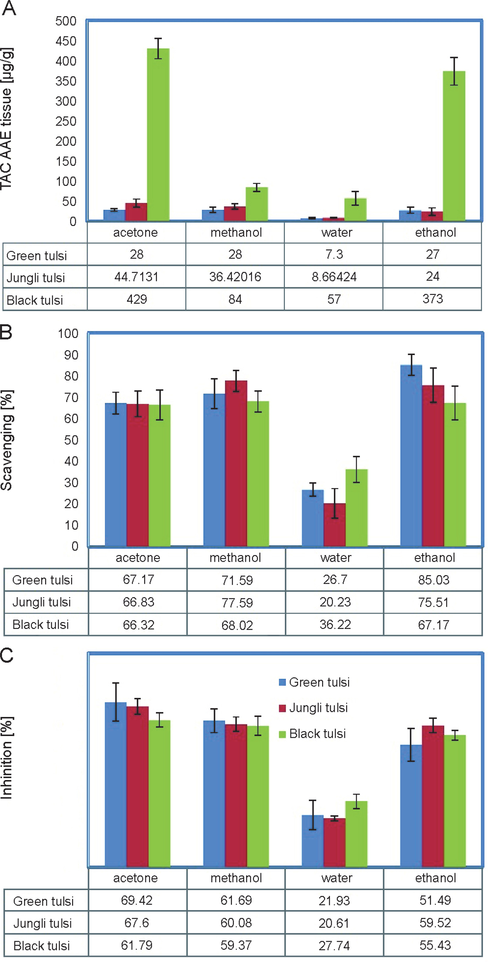
The antioxidant activity of medicinal plants is the key aspect used to reveal its therapeutic potential (Khan et al., 2012). Because plants contain a complex mixture of antioxidants, it is very difficult to measure the antioxidant capacity of individual components. Therefore, three methods based on 2,2-diphenyl-1-picrylhydrazy (DPPH) and total antioxidant activity measurements were used to evaluate the antioxidant potential of O. basilicum L. (Green tulsi), O. gratissimum L. (Jungli tulsi), and O. tenuiflorum (Black tulsi) extracts. The total antioxidant activity of all the three Ocimum species varied depending upon the solvent used. In the present study, acetone extracts of all Ocimum plants showed the maximum total antioxidant activity in the range of 28–429 ascorbic acid equivalent (AAE)/g DW, followed by the extracts in ethanol (24–373 AAE/g DW), methanol (28–84 AAE/g DW), and water (7–57 AAE/g DW) (Fig. 1A). Among all the three Ocimum spp., the extract of black tulsi in acetone showed the maximum total antioxidant activity of 429 AAE/g DW, followed by jungli tulsi (44 mg AAE/g DW) and green tulsi (28 mg AAE/g DW). Ethanolic extracts of all the Ocimum plants showed the maximum scavenging activity in the range of 67–85%, followed by the extracts in methanol (68–71%), acetone (66–67%), and water (20–36%) (Fig. 2B). Among all the three Ocimum species, green tulsi exhibited the maximum scavenging activity of 85%, followed by jungli tulsi (75%) and black tulsi (67%). Aggarwal et al. (2017) showed that the methanolic root extract of O. kilimandscharicum had better DPPH radical scavenging potential (12%), followed by the methanolic extract (6%) of O. sanctum. The authors claimed that the chloroform and ethyl acetate extracts of both the species exhibited better total anti-oxidant potential than the extracts in other solvents, wherein the values ranged from 7.56 ± 0.47 to 11.31 ± ± 1.02 μg AsE/mg of dried extracts. Khan et al. (2012) observed strong DPPH scavenging and antioxidant activities in the methanolic extracts of L. procumbens. Potent antioxidant DPPH scavenging activity has also been reported in the methanolic extracts of Synsepalum dulcificum (Obafemi et al., (2017). The therapeutic agents and herbal drugs that are mostly used to ameliorate free radicals and oxidative stress-related diseases are reported to have strong scavenging power (Khan et al., 2012). We therefore believe that the marked antioxidant activities of tulsi extracts may be attributed to the presence of phenolics, flavonoids, and other compounds as observed in the present study. Biological extracts with rich antioxidant activities have a great potential to be used as food supplement to control the damage of biomolecules by inhibiting the generation of free radicals in the biological system and consequently rejuvenating the body functions (Pattanayak et al., 2010). In the past few years, the identification of phenolic-based derivatives from different plant extracts has become an important area of health- and medical-related research (Mandave et al., 2014).
Fingerprint analysis
UV-vis, fluorescent, and FT-IR spectroscopy methods together or separately can be used as conventional methods for detecting the content of phytocomponents (Chen et al., 2016). Figure 4 shows the UV-vis profiles of all the three Ocimum plant extracts in acetone, ethanol, methanol, and water. The UV-vis spectroscopy profile of all the three tulsi extracts revealed absorption maxima at approximately 350 nm with an absorption range of 2.5–3.0. In the UV-vis profile, the occurrence of peaks in the region at 350 nm is a clear indication of the presence of polyphenolics in all tulsi plant extracts. These observations are consistent with the findings reported by Paulraj et al. (2011) in Christella parasitica extracts. Earlier studies also showed the presence of polyphenolics in herbal extracts of Cassia auriculata (Ranawara), Hemidesmus indicus (Iramusu), Aegle marmelos (Bael), and Aerva lanata (Polpala) (Chandrasekara and Fereidoon, 2018).
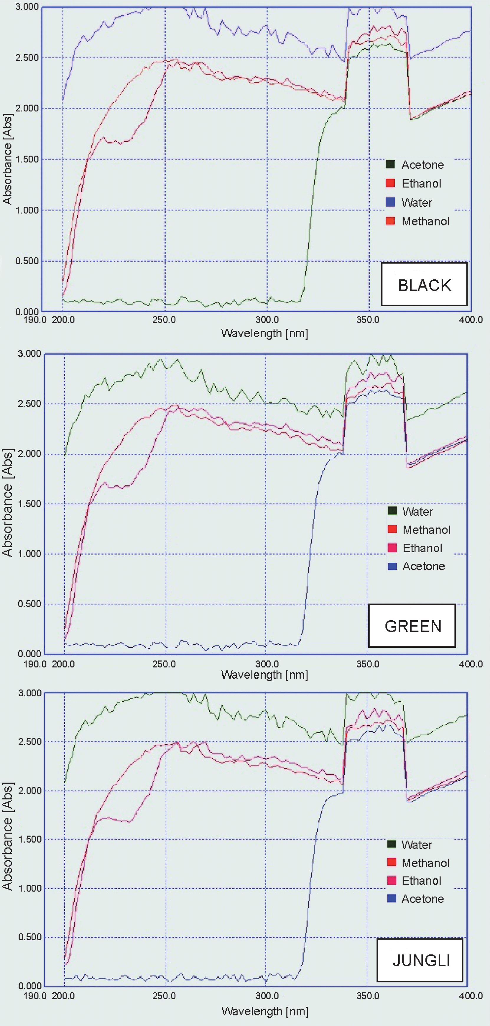
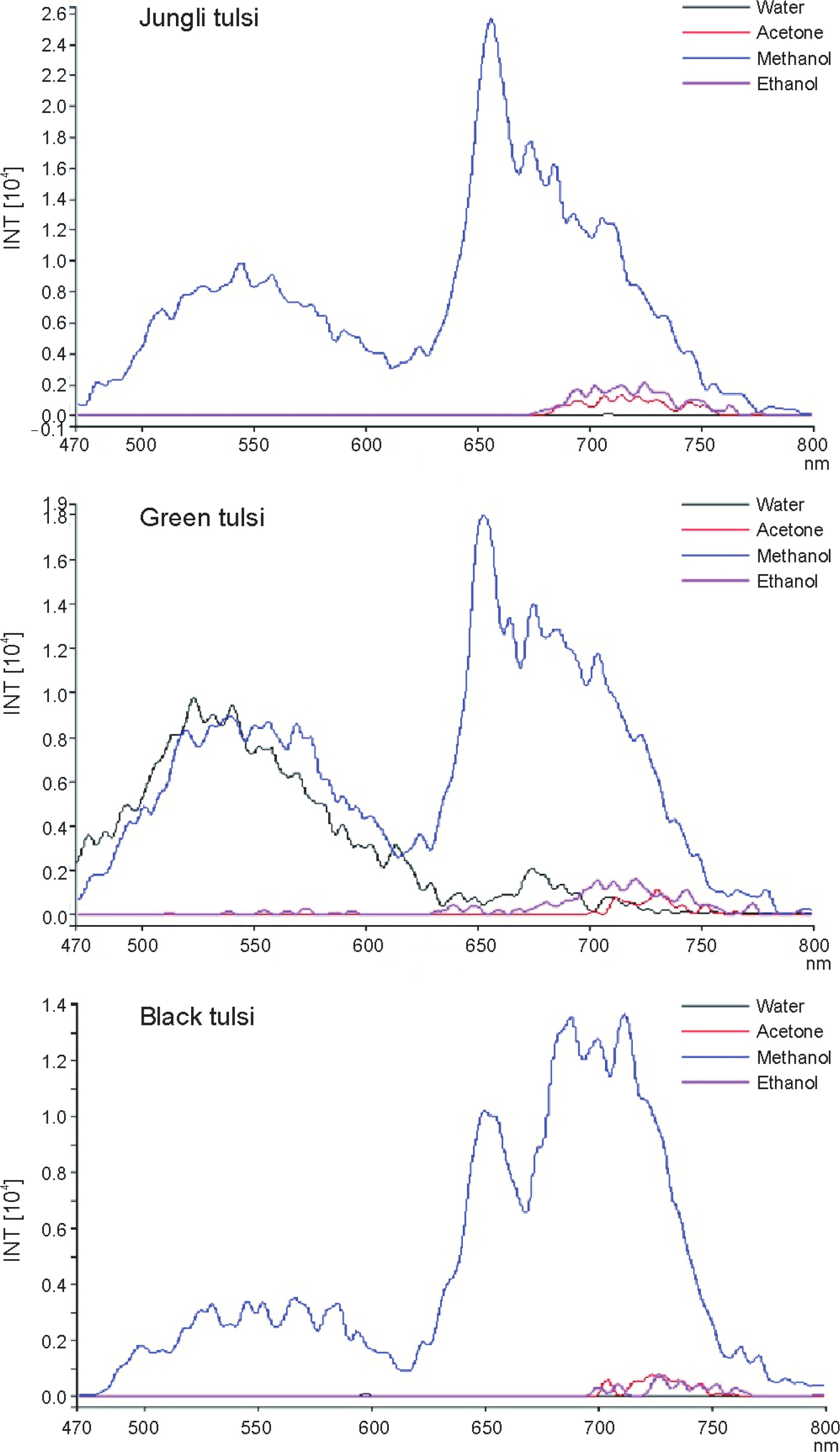
Figure 4 shows the fluorescent emission spectra of Ocimum spp. extracts. The extracts were grouped according to their emission spectra. Various bioactive components emit fluorescence when excited with a suitable light (Talamond et al., 2015). In jungli and black tulsi, two major peaks in the lambda region near 550 and 650 nm were detected only in methanolic extracts (Fig. 4), indicating that fluorescent substances are present in tulsi extract. Other solvents such as ethanol, acetone, and water were unable to extract a noticeable level of fluorescent compounds. In green tulsi, in addition to methanol, water could extract a sufficient amount of fluorescent molecules as observed by the presence of a sharp peak at 550 nm. The fluorescent candidates for the green emission region (lambda near 500 nm) are generally polyphenolics (Mylle et al., 2013, Talamond et al., 2015). Another set of bioactive compounds, as evidenced by a major peak around 650 nm, was also detected in the red florescent region (RF), which may be due to the accumulation of other fluorescent compounds such as anthocyanins, phenolics, alkaloids, and aromatic benzenoids (Mylle et al., 2013, Talamond et al., 2015).
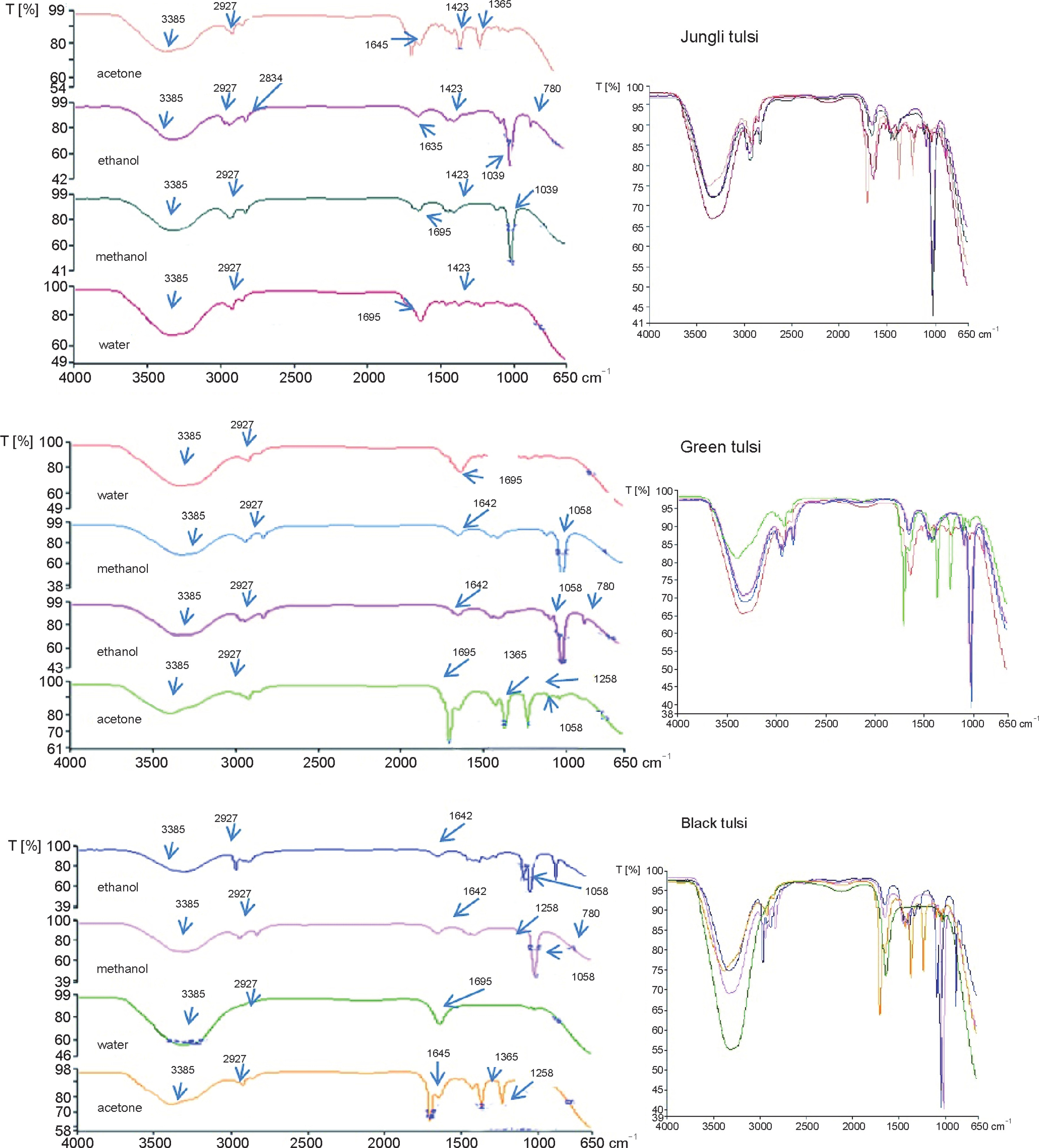
FT-IR spectroscopy has been used to detect the presence of different types of functional groups in active constituents of plant extracts (Chen et al., 2016). The FT-IR profile of tulsi extracts is shown in Figure 5. Table 1 shows the variations in FT-IR peaks. The FT-IR spectrum can be divided into four main regions: 1st: 4000–2500 cm−1, 2nd: 2500–2000 cm−1, 3rd: 2000–1500 cm−1, and 4th: 1500–400 cm−1. The 4th region is the major region of the IR spectrum, which is also known as the fingerprint region as it contains a large number of complex peaks (Türker-Kaya and Huck, 2017). All solvent extracts of the three Ocimum species depicted different FT-IR profiles. The ethanol and methanol extract profiles were, however, relatively similar. Each species exhibited its own characteristics such as different peak shapes, numbers, position, and intensity, thus indicating the presence of different functional groups. A broad band in the range of 3400 to 3200 cm–1 was observed in the FT-IR profile of all the three tulsi extracts. This band was attributed to the OH-group, which confirmed the presence of phenolic compounds in the extracts. Oliveira et al. (2016) also reported the presence of peaks at 3500 cm−1 due to O–H stretching of alcohols in Punica granatum. In extracts obtained from all the three Ocimum species in all the tested solvents, another sharp band at approximately 2800–2900 cm−1 was detected due to C–H stretching of alkanes. Peaks in the range of 1830–1650 cm−1 were detected in acetone, ethanol, and methanol extracts of all three Ocimum species, thus indicating the presence of aldehydes, ketones, esters, carboxylic acids, amides, and anhydrides. Within the same solvent, all the three Ocimum species showed almost the same FT-IR profiles with only minor differences. Differences were observed in the 4th fingerprint region in the range from 1500–1400 cm−1 as shown in the superimposed form of the FT-IR spectra (Fig. 5 and Table 1). For example, in jungli and black tulsi species, a peculiar band in the range of 1645 cm−1 was detected in acetone extracts, which was ascribed to C=C stretching of alkenes; this band was, however, absent in green tulsi. Notably, only in the water extracts of all the three Ocimum species, a peculiar band at 1695 cm−1 was observed, which was due to C=C stretching of alkenes. Jain et al. (2016) also ascribed peaks at around 1695 cm−1 to alkenes in Mentha spicata, while Bashyam et al. (2015) reported the same in Bryonopsis laciniosa. Oliveira et al. (2016) reported similar C=C stretching of alkenes in P. granatum. In acetone extracts of all the three Ocimum species, a peculiar band was detected in the range of 1365 cm−1. The band at 1365 cm−1 due to bending (δ) vibration of C–H could be related to CH3 , CH2, flavonoids, and aromatic rings as indicated by Silva et al. (2014) in Propolis and Bashyam et al. (2015) in Bryonopsis laciniosa. In the range of 1150–1021 cm−1, minor bands due to C–N stretching of aliphatic amines were detected in the ethanolic and methanolic extracts of all the three Ocimum species. One peculiar band in the range of 780 cm−1, due to 1,2,3-tridistribution, was detected only in the ethanolic extracts of all the three Ocimum species. Peaks at approximately 1362 cm−1 were due to carboxylic acids, esters, and ethers and those at 1092 cm−1 could be due to C–O stretching and –OH de-formation of secondary alcohols as reported by Oliveira et al. (2016) in P. granatum and Bashyam et al. (2015) in B. laciniosa. All these compounds belong to secondary plant metabolites (Paulraj et al., 2011). These observations imply that all Ocimum species are rich in secondary metabolites, but their activity varies in a solvent-dependent manner. The presence of the above-mentioned secondary metabolites may be the reason for medicinal properties of the tulsi plant.
Table 1
FT-IR peaks in all Ocimum species extracts tested
Antidiabetic activity of Ocimum extracts
Inhibitors of α-amylase delay the conversion of carbohydrates in the small intestine and lower the level of prandial blood glucose surge in persons with diabetes (Toma et al., 2015). Many therapeutic strategies involve the inhibition of carbohydrate-digesting enzymes such as α-amylase to treat diabetes mellitus. In the present study, the effects of Ocimum species plant extracts on α-amylase activity were assessed. The extracts of all the three Ocimum species showed dramatic inhibition of α-amylase activity in a solvent-dependent manner (Fig. 2C).
In all the three Ocimum species, the maximum antidiabetic activity was observed in the acetone extract (61–69% of α-amylase inhibition), followed methanol (59–60%) and ethanol (51–59%) extracts. In acetone extracts, green tulsi showed the maximum level of antidiabetic activity with 69% inhibition, followed by jungli tulsi (67% inhibition) and black tulsi (61% inhibition). In acetone and methanol extracts, almost the same level of antidiabetic activity was observed in all the three Ocimum species. The water extracts of the three species showed the lowest antidiabetic activity in the range of 20–27% inhibition. Parasuraman et al. (2015) observed 50% antidiabetic activities in the hydroalcoholic extract of O. tenuiflorum. Suanarunsawat et al. (2016) also reported 40–50% antidiabetic activity in the hexane extract of O. sanctum. These results agree with previous reports that highlighted the inhibition of α-amylase activity by medicinal plant extracts (Kwon et al., 2008). The results of the present study suggest that tulsi extracts have hypoglycemic potential, which may be ascribed to the presence of flavonoids and phenolics. An earlier study on alpha-amylase inhibitors from medicinal plants suggest that several potential inhibitors belong to the group of flavonoids (Mccue et al., 2005).
Anti-inflammatory activity of Ocimum species extracts
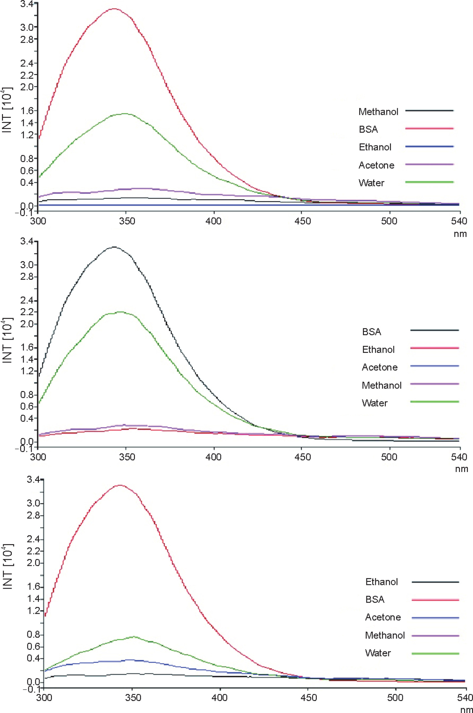
Protein denaturation is the process in which native proteins lose their tertiary and secondary structures. The major reason underlying the loss of protein functions is the disruption of hydrogen, hydrophobic, and disulfide bonds in protein structures (Sridevi et al., 2015). Protein denaturation is a well-established cause of tissue inflammation that is associated with symptoms such as redness, pain, heat, and swelling (Kumar et al., 2018). Hence, it was deduced that compounds that can prevent these changes and inhibit heat-induced protein denaturation have the potential to be used as therapeutic anti-inflammatory drugs (Dharsana and Mathew, 2014). In the present study, the potential of Ocimum species plant extracts to inhibit protein denaturation was investigated by tryptophan fluorescent spectroscopy (Sharma et al., 2021). Typical fluorescence spectra for denatured bovine serum albumin (BSA) and the Ocimum species plant extracts in different solvents are shown in Figure 3. The acetone, methanol, and ethanol extracts of all the three Ocimum species protected BSA against protein denaturation, as fluorescence intensity decreased considerably from 3.4 × 10⁴ INT units to 0.2 × 10⁴ INT units after the addition of the extracts to BSA. Among all the extracts, the maximum anti-inflammatory activity was observed for the ethanolic extract, with approximately 3-fold decrease in fluorescence intensity as compared to that for other solvents. The least protection of BSA against denaturation was shown by the water extract of jungli tulsi (decrease in fluorescence intensity from 3.4 × 10⁴ to 1.4 × 10⁴ INT units) and green tulsi (decrease in fluorescence intensity from 3.4 × 10⁴ to 2.2 × 10⁴ INT units). The water extract of black tulsi, however, significantly prevented BSA denaturation, with a sharp decrease in fluorescence intensity from 3.4 × 10⁴ to 0.6 × 10⁴ INT units. These results clearly indicate the potential of Ocimum species extracts as anti-inflammatory agents. The anti-denaturation activity may be ascribed to complex mixtures with multiple compounds that may interact synergistically. Tatti et al. (2012) observed substantial in vitro anti-inflammatory activity in the ethanolic extracts of Butea monosperma. Dharsana and Mathew (2014) reported in vitro anti-inflammatory activity in ethanolic extracts of Morinda umbellata. Kumar et al. (2018) demonstrated potent in vitro antiinflammatory activity in the hydroethanolic extracts of Celastrus paniculatus.
Conclusions
The present study evaluated the phytochemical and biological potential of various solvent extracts of Ocimum species. The extracts of all the three Ocimum species, especially black tulsi, contained high levels of secondary metabolites such as phenolics, flavonoids, and tannins and showed high antioxidant activity. Strong anti-diabetic and anti-inflammatory activities were also observed in the ethanolic and acetone extracts of the three Ocimum species. It was noted that the selection of an appropriate solvent that could extract sufficient levels of bioactive compounds from the plants could enable to provide high biological activities in vitro. The high content of secondary metabolites in the Ocimum species plant extracts can be used as a reference source of antioxidants in food and pharmaceutical-based industries.